
 This work is licensed under a
 Figure 1.
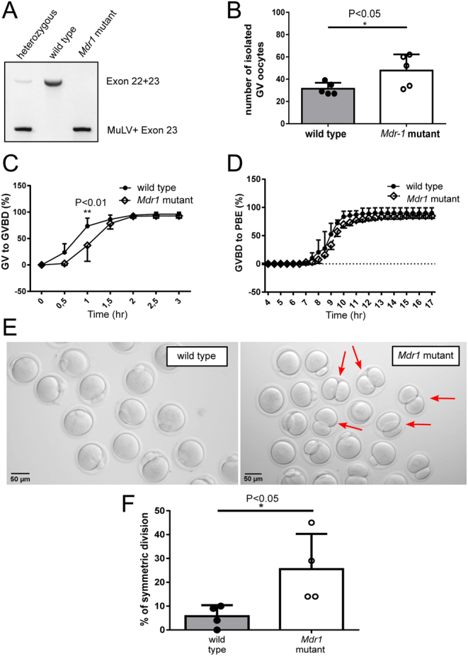
Two-month-old mutant oocytes show a delayed germinal vesicle breakdown (GVBD), and the resulting MII oocytes present enlarged first polar bodies (PBs). (A) Mouse genotyping. MuLV, murine leukemia virus insertion. (B) Number of GV oocytes isolated from wild-type and Mdr1 mutant mice. Unpaired t-test; *P < 0.05. n = 5 wild-type and 5 Mdr1 mutant mice. (C) Percentage of young mutant and wild-type oocytes transitioning from germinal vesicle to germinal vesicle breakdown (GVBD) 30 min after experiment start and 60 min after milrinone removal. Both wild-type and mutant oocytes reach GVBD, although mutant oocytes are significantly delayed in the transition. Two-way ANOVA; **P < 0.01. (D) Percentage of oocytes extruding the first polar body (PBI) after 16 h of in vitro maturation. No difference was observed in the frequency and timing of polar body extrusion (PBE) between wild-type and mutant oocytes. n = 3 wild-type and 3 Mdr1 mutant mice. (E and F) Images representing the different PB sizes in wild-type and mutant oocytes. Oocytes with symmetric divisions are indicated by a red arrow. Unpaired t-test; *P < 0.05. Scale bar = 50 µm. n = 3 wild-type and 3 Mdr1 mutant mice.