Figure 2.
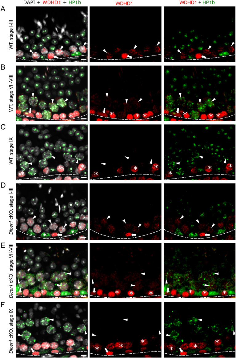
WDHD1 is expressed by spermatogonia and early spermatocytes in WT and Dicer1 cKO mice. (A) In stage I–III of WT mouse seminiferous epithelial cycle, WDHD1 (red) is highly expressed in spermatogonia (arrow), whereas the expression level in early-pachytene spermatocytes (arrowhead) is low. HP1β (green) stains heterochromatin. (B) In WT stages VII–VIII, WDHD1 is expressed by spermatogonia (arrow) and preleptotene spermatocytes (asterisk). In mid-pachytene spermatocytes (arrowhead), the WDHD1 expression level is low. (C) By WT stage IX, late-pachytene spermatocytes (arrowhead) become negative for WDHD1. WDHD1 is also downregulated upon preleptotene-to-leptotene (asterisk) transition. In Dicer1 cKO mice, the expression pattern of WDHD1 is similar to WT in stages (D) I–III, (E) VII–VIII and (F) IX. DAPI stains chromatin (white). Basement membrane of seminiferous epithelium (dotted line), spermatogonia (arrow), early spermatocytes (asterisk), pachytene spermatocytes (arrowhead). Scale bars 10 µm.

 This work is licensed under a