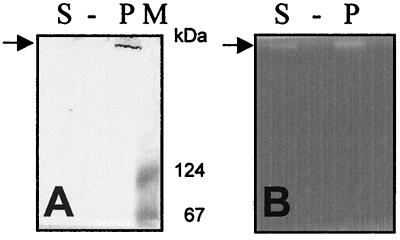
FIG. 3.
SpuA functional analysis. The same polyacrylamide gel in which the S. pneumoniae strain 3.B native whole-cell extract (P) and the culture supernatant (S) were separated was used for Western blot analysis (A) and detection of pullulanase activity (B). The protein band recognized by anti-SpuA and possessing pullulanase activity is indicated by arrows. Lane M, molecular mass standard (sizes shown at right).

