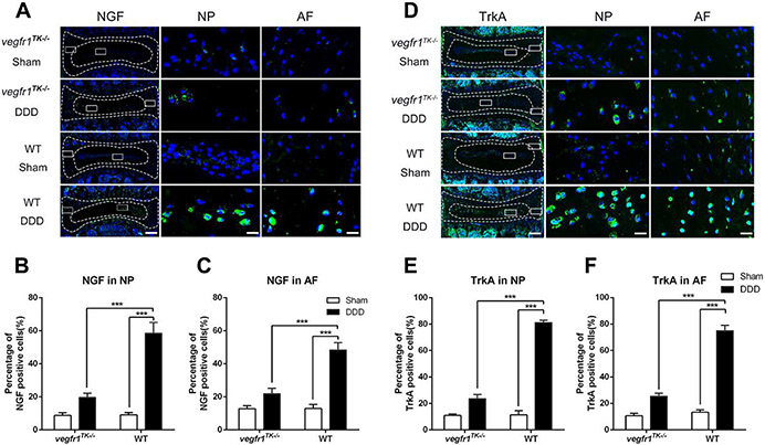
Figure 3.
NGF and TrkA expression was reduced in the discs of vegfr-1TK−/− mice compared to WT mice of the DDD groups. Representative immunofluorescence staining for NGF (A) and TrkA (D) in the IVD of DDD and sham mice at 12 weeks post-surgery (bars=200μm for the integral discs, bars=20μm for magnified NP and AF areas). Quantitative analyses showed that NGF (B, C) and TrkA (E, F) expression in IVDs of the DDD group of vegfr-1TK−/− mice was significantly lower than that in the IVDs of the DDD group of wt mice at 12 weeks after surgery. (n=5 per group) (***P< 0.001).

