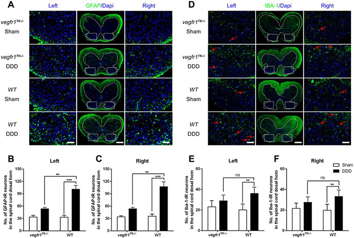
Figure 5.
Astrocyte and microglia reactivity in the lumbar part of the spinal cord following disc stabbing surgery. (A & D) Representative immunofluorescence images showing results of staining for glial fibrillary acidic protein (GFAP, an astrocyte marker) (A) and ionized calcium–binding adapter molecule 1 (IBA-1, a microglia marker) (B) in lumbar spinal cord of each group of mice(bars=100μm for the integral spinal cord and bars=20μm for the magnified dorsal horn areas). (B & C) Quantitative analyses show that at 12 weeks post-surgery, although the number of GFAP-ir neurons in both DDD group were increased compared to the respective sham group, the number in the DDD group of vegfr-1TK−/− mice were significantly lower than that in the DDD group of wt mice. (E & F) The number of IBA-1-ir neurons(red arrows) in the DDD group of wt mice markedly increased compared to the respective sham group, while there was no difference between the DDD group and the sham group of vegfr-1TK−/− mice and no difference between both DDD groups of vegfr-1TK−/− and WT mice. (n=5 per group). (**P< 0.01, ***P< 0.001; ns, no significance)

