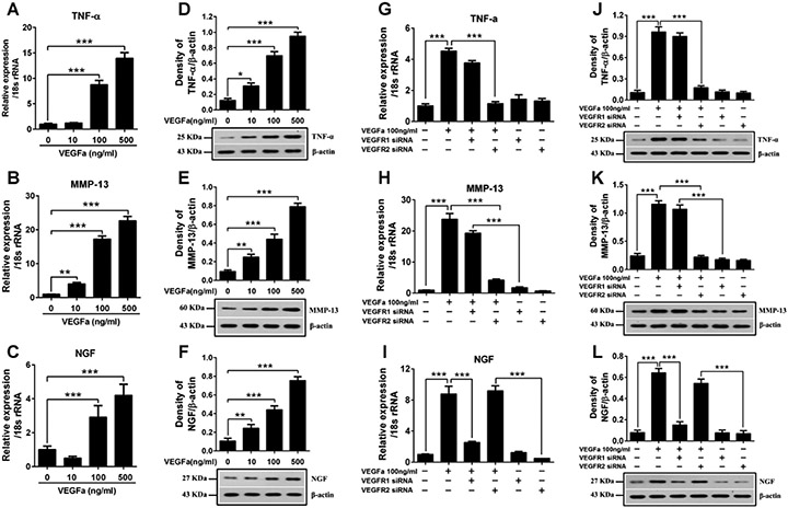
Figure 6.
Analysis of different functions of VEGF mediated by VEGFR-1 and VEGFR-2 on bovine NP cells. (A-F) VEGF-A at different concentration was added to the NP cells and incubated for 24 hours after starving in serum-free medium over night. Real-time PCR and western blot analyses revealed that introduction of VEGF-A to bovine NP cells significantly increased the expression of TNF-α, MMP-13 and NGF in a dose-dependent manner for RNA levels (A-C) and protein levels (D-F). (G-L) Bovine NP cells were transfected with VEGFR-1 siRNA or VEGFR-2 siRNA with Lipofectamine 2000 and then treated with VEGF-A 100ng/ml for 24 hours. Real-time PCR and western blot analyses revealed that VEGFR-1 silencing significantly suppressed the VEGF-A induced over expression of NGF (I, L), while the expression of TNF-α (G, J) and MMP-13 (H, K) was still significantly increased compared to the negative control. And VEGFR-2 silencing markedly reduced the VEGF-A induced over expression of TNF-α (G, J) and MMP-13 (H, K), while the expression of NGF (I, L) still remained at a significantly higher level. All values are mean ± SEM. *P<0.05, **P<0.01, ***P< 0.001.

