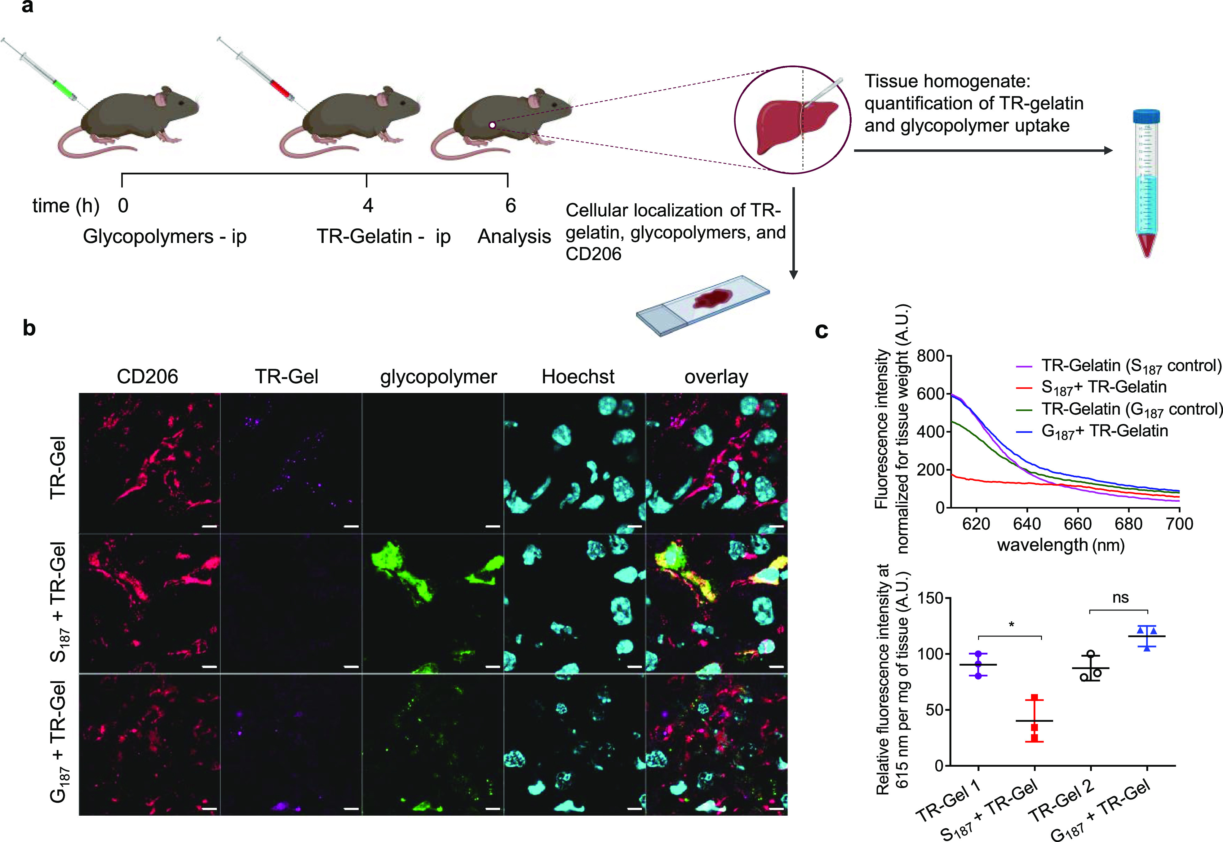
Figure 7.

S187 inhibits uptake of gelatin by liver cells in vivo. (a) Mice were injected intraperitoneally with 100 μL of Oregon Green (OG)-tagged S187 or G187 (490 μM polymer units in PBS). Four hours later, mice received 100 μL of TR-gelatin solution (1.0 mg mL–1 in PBS), and livers were collected after further 2 h. Mice treated with TR-gelatin for 2 h were used as positive controls. Created with BioRender. (b) Immunofluorescence analysis of liver sections: representative two-dimensional fluorescence images obtained by confocal laser scanning with Hoechst staining for nuclei (cyano), immunostaining for the CD206 receptor (red), TR-gelatin (magenta), and S187 or G187 glycopolymers (green). Mice were treated with TR-gelatin only (TR-Gel) or S187 or G187 followed by TR-gelatin (S187 + TR-gelatin or G187 + TR-gelatin, respectively) as described in the Materials section. Scale bars: 5 μm. Series of 0.5 and 2× magnification images are shown in Figures S15–S17; and (c) liver TR-gelatin uptake was quantified on liver homogenate as relative fluorescence signal at λem = 615 nm per mg of liver tissue homogenate. Top panel: representative emission spectra in the 610 to 700 nm range (λex = 596 nm for TR-gelatin quantification) from a single animal. Bottom panel, collated data λem = 615 nm from N = 3. TR-Gel 1 and TR-Gel 2 are the positive control samples (mice not pre-treated with glycopolymers) for S187 + TR-Gel and G187 + TR-Gel samples, respectively. A two-tailed t-test was performed to test significance; * P ≤ 0.05.
