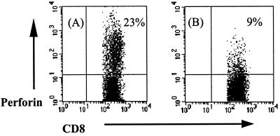
FIG. 1.
Flow cytometric detection of intracellular perforin by CD8+ T cells. PBMC from a healthy BCG vaccinee (A) and a pulmonary TB patient (B) were stimulated for 7 days in the presence of live M. tuberculosis. For the final 16 h of stimulation, brefeldin A was added to the cultures. Intracellular perforin staining was performed before FACS analysis. Cells were progressively gated by forward- and side-scatter for lymphocytes, and analysis was performed on the CD8+ high-staining lymphocytes. Results shown are for one BCG vaccinee and one TB patient who are representative of the 10 donors in each group tested.

