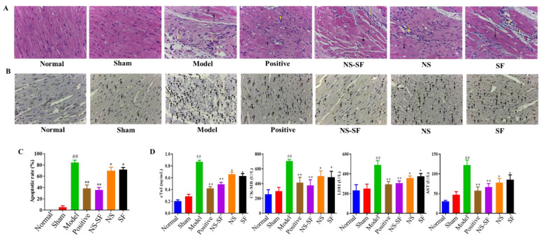
Figure 2.
Effects of NS, SF and NS–SF on myocardial fibrosis and apoptosis induced by MI in rat heart tissues. (A) H&E staining of left ventricular (LV) tissue showed pathological and morphological changes in different groups (magnification, ×400). The necrotic myocardium is labeled with a black arrow, and inflammatory cells are labeled with a yellow arrow. (B) The results of TUNEL staining in different groups. Arrows indicate apoptotic cardiomyocyte nuclei. (C) Apoptosis rate of myocardial cells in each group (mean ± SD, n = 6). (D) The serum levels of MI-associated biochemical markers of rats in different groups (mean ± SD, n = 6). ## p < 0.01, when versus sham group; * p < 0.05, ** p < 0.01, when versus model group. Normal: normal group; Sham: sham-operated group; Model: model group; Positive group: positive control group; NS: total saponins of notoginseng group; SF: total flavonoids of safflower group; NS–SF: the compatibility of NS and SF group.

