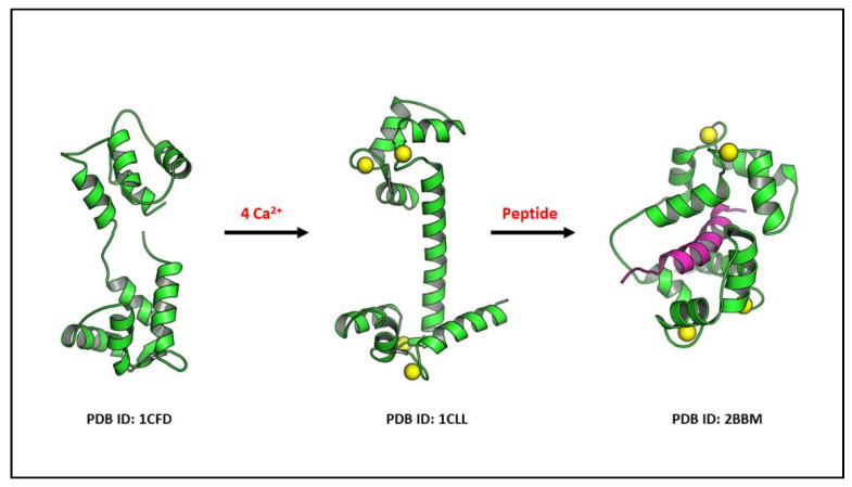
Figure 2.
The 3D illustrations show the flexibility of calmodulin in binding to 4 Ca2+ and undergoing conformational changes for its reaction with a specific downstream protein, such as the CaM kinase proteins. Specifically, the binding of the calcium–calmodulin complex to auto-inhibitory target protein regions leads this conformational change and the target protein activation. Ca2+ is displayed as yellow balls and peptide is shown as magenta ribbon.

