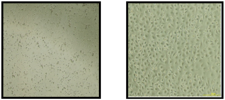
Figure 4.
Morphological microscopic appearance of monocytes and macrophages. The figure on the left represents the shape of the isolated monocyte cells under the light microscope (×10) at day 1 immediately after magnetic separation. The figure on the right shows the monocyte-derived macrophages under the microscope (×10) after 7 days of monocyte culturing in the presence of GM-CSF. GM-CSF, granulocyte-macrophage colony stimulating factor.

