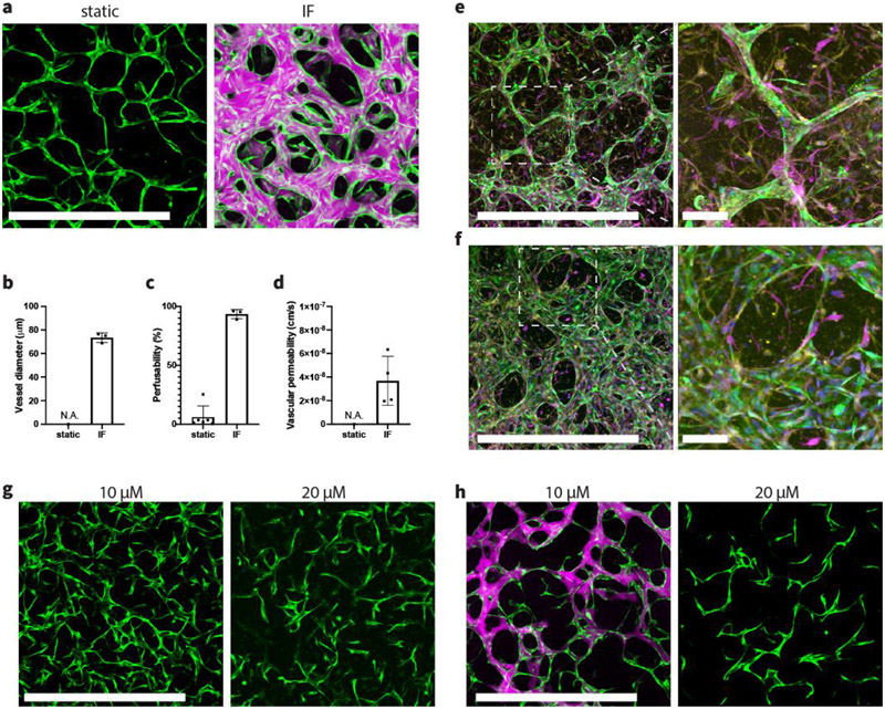
Figure 6.
IF enhances formation of fully perfusable brain MVNs. (a) Brain specific MVNs formed statically or under IF induced by 10 mm H2O pressure difference. Green – GFP BECs, magenta – 40 kDa Texas Red dextran. Scale bar is 1 mm. (b,c) Statistical analysis of vessel diameter and perfusability of brain MVNs formed statically or under IF. n=6 devices for static group; n=3 devices for IF group. (d) Vascular permeability measurements using 40 kDa dextran. n=2 devices, 2 measurements in each device at different ROIs. (e) Fluorescent confocal images of brain specific MVNs cultured statically at day 6 with zoomed-in view. (f) Fluorescent confocal images of brain specific MVN cultured under IF induced by 10 mm H2O pressure difference at day 6, with zoomed-in view. Green – GFP BECs, magenta – S-100b staining for ACs, yellow – PDGFR staining for PCs. Scale bar is 1 mm for the left panel, and 100 μm for expanded view. (g) Day 3 brain MVNs cultured under IF, supplemented with 10 μM or 20 μM ARP-100. Scale bar is 1 mm. (h) Day 6 brain MVNs cultured under IF, supplemented with 10 μM or 20 μM ARP-100. Green – GFP BECs, magenta – 40 kDa Texas Red dextran. Scale bar is 1 mm.

