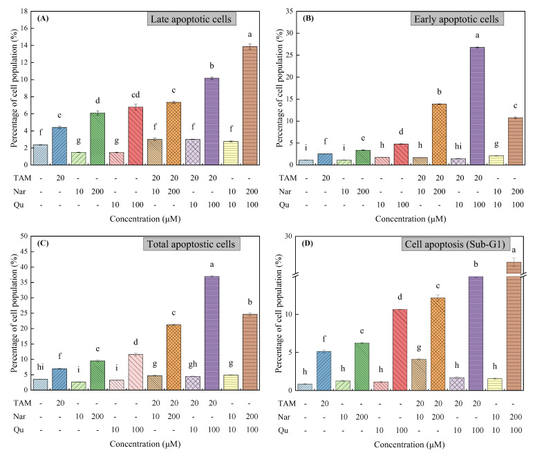
Figure 5.
Tamoxifen, naringenin and quercetin regulated cell apoptosis in HepG2 cells. Cells were exposed to tamoxifen (20 μM), naringenin (10 and 200 μM) and quercetin (10 and 100 μM) either alone or in combination for 24 h. (A–C) Percentage of cell population in the different apoptotic stages. (D) Sub-G1 DNA content of cells analyzed with PI staining. Results are expressed as mean ± SD of three independent experiments (n = 3). a–i: Different letters indicate significant differences (p < 0.05) in cell apoptosis.

