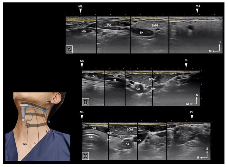
Figure 5.
The ultrasonographic image of the platysma muscle was observed in blue arrow direction. The muscle lies from the midline to the vertical line from the mandibular angle and has been presented in the ultrasonographic image of three transverse planes: below the mandibular border (A), superior notch of the thyroid cartilage (B), and cricoid cartilage (C). (ML, midline; MH, Mylohyoid muscle; DG, Digastric muscle; SH, Sternohyoid muscle; SMG, Submandibular gland; MA, mandibular angle; CA, carotid artery; OHS, Omohyoid superior belly; OHI, Omohyoid inferior belly; IJV, Internal jugular vein; TH; Thyrohyiod muscle; TG; Thyroid gland; CC, Cricoid cartilage; IL, intermediate line; LL, lateral border of the lateral band).

