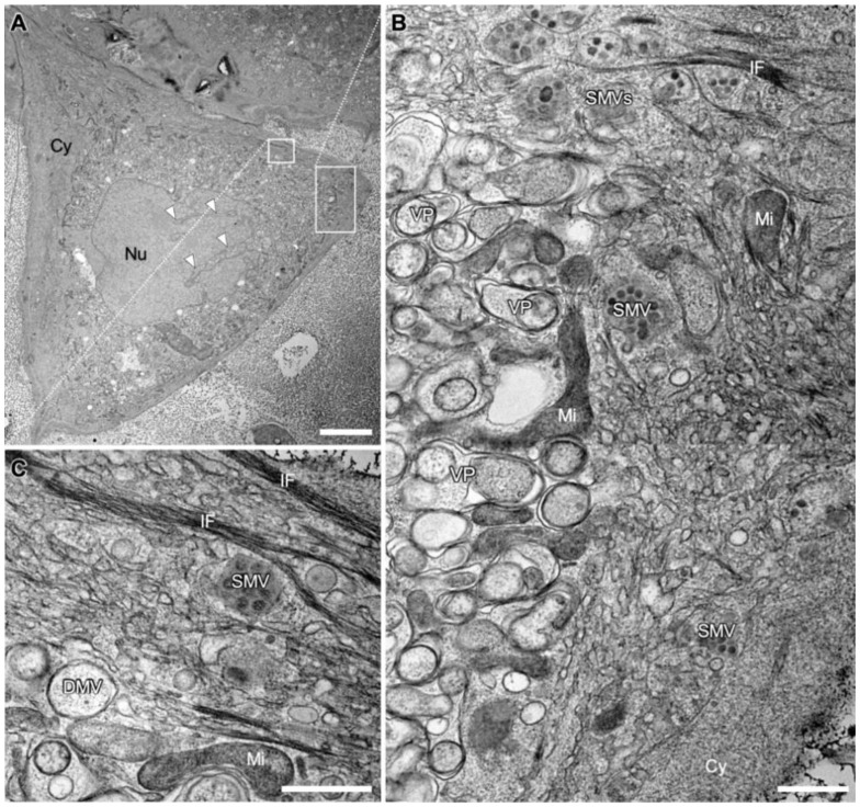
Figure 1.
Transmission electron microscopy (TEM) images of a SARS-CoV-2 infected Vero E6 cell at 24 hpi. (A) Overview image showing cytopathic alterations, such as the compartmentation of the cytoplasm (Cy). Invagination of the cytoplasm into the nucleus (Nu) is marked with white arrowheads. (B) Larger magnification of the indicated area marked in A, illustrating the compartmentation into a perinuclear area on the left and a peripheral area on the right. The perinuclear area is occupied almost exclusively by densely packed double membrane vesicles (DMVs) and numerous vesicle packets (VPs), arising from multiple DMVs fusing at their outer membrane [19,24]. The peripheral area is occupied by cell organelles such as mitochondria (Mi) and endoplasmic reticulum (ER) and single membrane vesicles (SMVs) filled with virions. (C) Larger magnification of the area indicated in A with bundles of intermediate filaments (IF), SMV, DMV and a mitochondrion (Mi). Scale bars: (A) 10 µm; (B,C) 500 nm.

