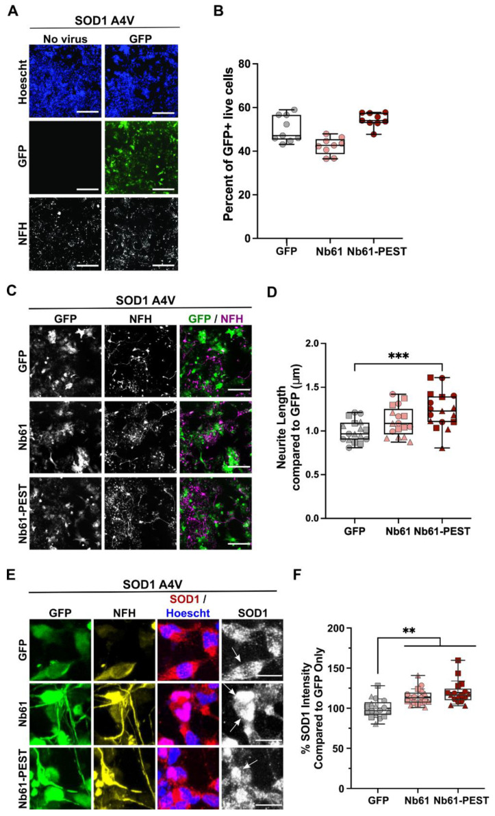
Figure 6.
Nb61-PEST promotes neurite outgrowth in human SOD1 A4V motor neurons. SOD1 A4V iPSC-derived human motor neurons were thawed, plated into 384-well dishes, and assessed under various conditions after 7 days in culture. (A) Immunofluorescence images of human SOD1 A4V neurons that were untreated (left) or treated (right) with a control lentivirus expressing GFP. Neurofilament heavy (NFH, SMI32) staining identified motor neurons. Scale bar = 200 μm. (B) Quantification of the transduction efficiency for a control lentivirus expressing GFP or lentiviral constructs expressing either Nb61 or Nb61-PEST with a GFP reporter. Data are compiled from multiple wells (each point represents one well) from a representative biological replicate. (C) Immunofluorescence images of SOD1 A4V neurons that were transduced with the indicated lentivirus and stained as in (A). Scale bar = 200 μm. (D) Quantification of total neurite length for SOD1 A4V neurons transduced with the indicated lentivirus revealed significantly enhanced neurite outgrowth upon expression of Nb61-PEST compared to the GFP control virus. Data are compiled from n = 3 biological replicates; each replicate is denoted by a distinct symbol. (E) As in (C) with additional anti-SOD1 staining. Scale bar = 25 μm. Arrows indicate cells that are GFP+, NFH+, and have a clear SOD1 signal. (F) Quantification of endogenous SOD1 fluorescence signal intensity from images shown in (E) demonstrates that transduced Nb61 and Nb61-PEST lead to enhanced SOD1 expression in SOD1 A4V neurons. Data are pooled across n = 3 biological replicates, with each point representing data acquired within a single well. For D and F, statistical analyses were performed with the Kruskal–Wallis test and Dunn’s multiple comparison test; ** p < 0.01, *** p < 0.001.

