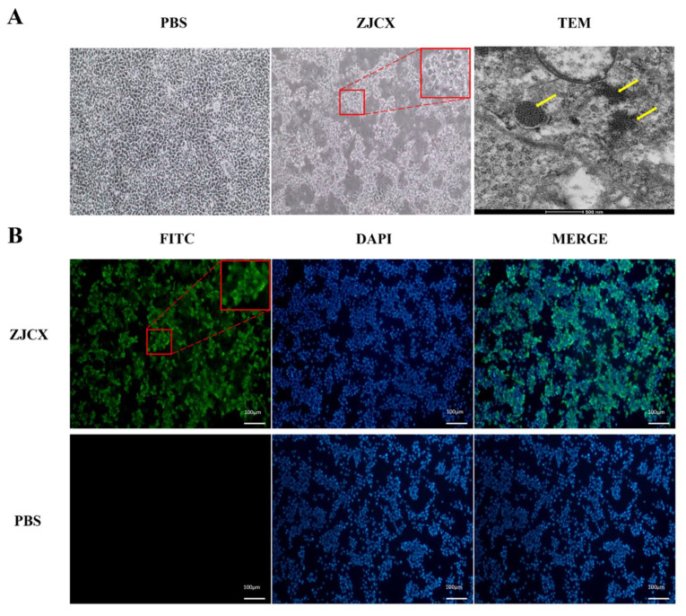
Figure 2.
Isolation of GAstV in LMH cells and identification of the GAstV ZJCX isolate by indirect immunofluorescence assay. (A) LMH cells with PBS inoculation as a negative control. The ZJCX inoculation could cause CPE in LMH cells (the red magnification was 2-fold, X2) and transmission electron micrograph of ZJCX in cells displaying a crystalline arrangement (yellow arrow). (B) LMH cells infected with ZJCX were reacted with rabbit serum against the capsid protein at 48 hpi, and a secondary antibody labeled with FITC displayed a green signal (the red magnification was 2-fold, X2). PBS as the negative group only displayed a blue signal, which was stained with DAPI for the cell nucleus.

