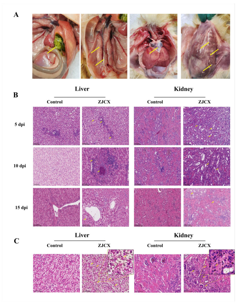
Figure 5.
Postmortem and histopathology lesions of tissues from goslings infected with GAstV. The dead goslings at 7 dpi displayed significant visceral gout with urate deposition (yellow arrow). Urate deposits in the gallbladder, ureters, peritoneum, pericardium, severe hemorrhage, and swellings of kidneys (A). HE-stained liver and kidney section with experiment goslings 5, 10, and 15 dpi showed necrosis, diffuse hemorrhage, renal tubular epithelial cell degeneration, and exfoliation. The histopathology change was shown with a yellow arrow. Magnification, ×200 (B). GMS stain of liver and kidney tissues from infected goslings 5 dpi showed urate with black color (yellow arrow) and deposits in the tissue (C).

