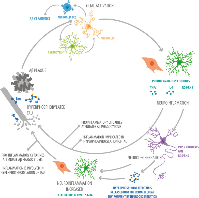
Fig. 1.
Microglial M2 and M1 phenotypes during AD. Two microglial activation phenotypes fluctuate in AD: M1, the classic and inflammatory phenotype (associated with release of ROS, RNS, and proinflammatory cytokines such as TNF-α and IL-1β); and M2, the alternative and anti-inflammatory phenotype (associated with release of anti-inflammatory cytokines such as IL10 and IGF1), which is responsible for Aβ phagocytosis. M1-to-M2 polarization is stimulated by proinflammatory cytokines such as IL4 and IL13, while M2-to-M1 polarization is stimulated by a lack of anti-inflammatory cytokines such as IL10 and IGF1. The M1 phenotype is seen in earlier stages of the disease, while the M2 phenotype is seen later. An example of a receptor involved in M2 activation is the nuclear receptor PPARγ, and an example of a pathway involved in M2 activation is the LKB1-AMPK pathway. An example of a receptor involved in M2 activation is the TLR2 receptor, which is capable of binding to Aβ, and an example of a pathway involved in M2 activation is the MYD88 pathway

