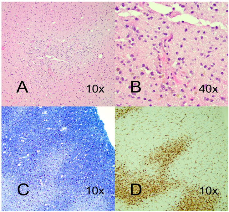
Figure 3.
Hematoxylin and eosin (HE)—stained section from right temporal lobe biopsy demonstrated perivascular infiltrates of macrophages and small CD3+ lymphocytes (A) with hemosiderin deposits as a sign of former hemorrhage (B). Luxol fast blue stain showed perivascular demyelination (C), and CD68+ stained macrophage infiltrates (D).

