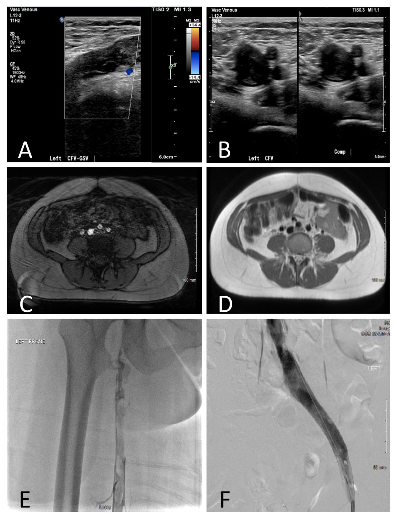
Figure 2.
Diagnostic Imaging from Patient 1. Duplex/Doppler ultrasound demonstrating occlusive thrombus in the CFV-GSV (A). Coronal T1-weighted (B) and axial T2-weighed MRA/V (C) demonstrating compression of the LCIV between the spine and RCIA, with extensive thrombus (arrows). IVUS demonstrating occlusive thrombus (D). Venography before (E) and after (F) stent placement postpartum.

