Figure 7.
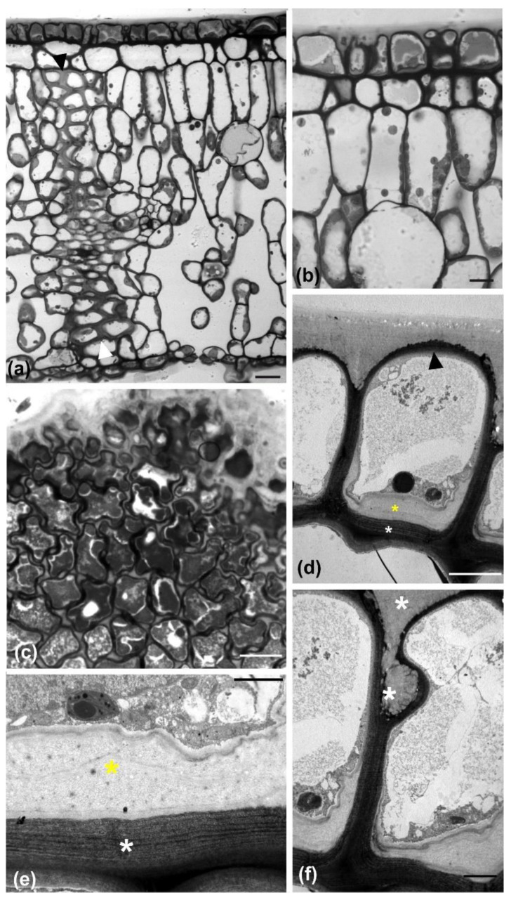
Light (a–c) and TEM (d–f) micrographs of mature leaves at transverse (a,b,d–f) or paradermal (c) section. At low magnification of transverse section (a), a “wall” of sclerenchyma cells can be observed (arrowheads) extending between the adaxial and abaxial epidermides. In addition, the external adaxial epidermal cells appear partially detached, which is shown at higher magnification in (b). The detachment is also visible at grazing paradermal section (c). (d) At TEM level, external adaxial epidermal cells exhibit a polylamellate inner periclinal wall (white asterisk), thicker than the external periclinal one (arrowhead), lined by the deposition of polysaccharide material (yellow asterisk). (e) Higher magnification of an inner periclinal wall area, like that in (d), with same labeling. (f) Higher magnification of a detachment site between two adjacent adaxial epidermal cells, depicting the stuffing with cuticle material (asterisks). Scale bars a: 20 μm, b, d: 5 μm, c: 10 μm e, f: 2 μm.

