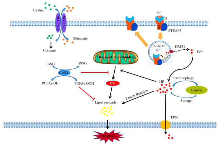
Figure 1.
Schematic diagram illustrating the proposed mechanism of ferroptosis. Figure legend: The occurrence and regulatory mechanisms of ferroptosis in a cell. Fe3+, which is indispensable to ferroptosis, can bind with Tf to form a Tf–Fe3+ complex and is internalized in cells by TFR using endocytosis. Then, Fe3+ is dissociated from Tf in an acidic environment and reduced to Fe2+ by STEAP3. Subsequently, Fe2+ is released from the endosome into LIP in the cytoplasm via DMT1. Free Fe2+ further results in lipid peroxide through the Fenton reaction, which ultimately triggers the occurrence of ferroptosis. In addition, some ferroptosis inducers can inhibit system xc- and impede the uptake of cystine by cells, thus leading to a decline in intracellular cysteine and a subsequent reduction in GSH, which requires cysteine for its synthesis; this ultimately results in a decline in the anti-oxidative ability of cells. As a key component in ferroptosis, GPx4 can bind with GSH and suppress cellular lipid peroxides to prevent cellular ferroptosis. Meanwhile, some substances can directly suppress GPx4 to induce ferroptosis. Mitochondria are the most important organelle involved in ferroptosis, which are the main sites for the generation of ROS and release of ferroptosis-inducing lipid peroxides, especially when they are damaged.

