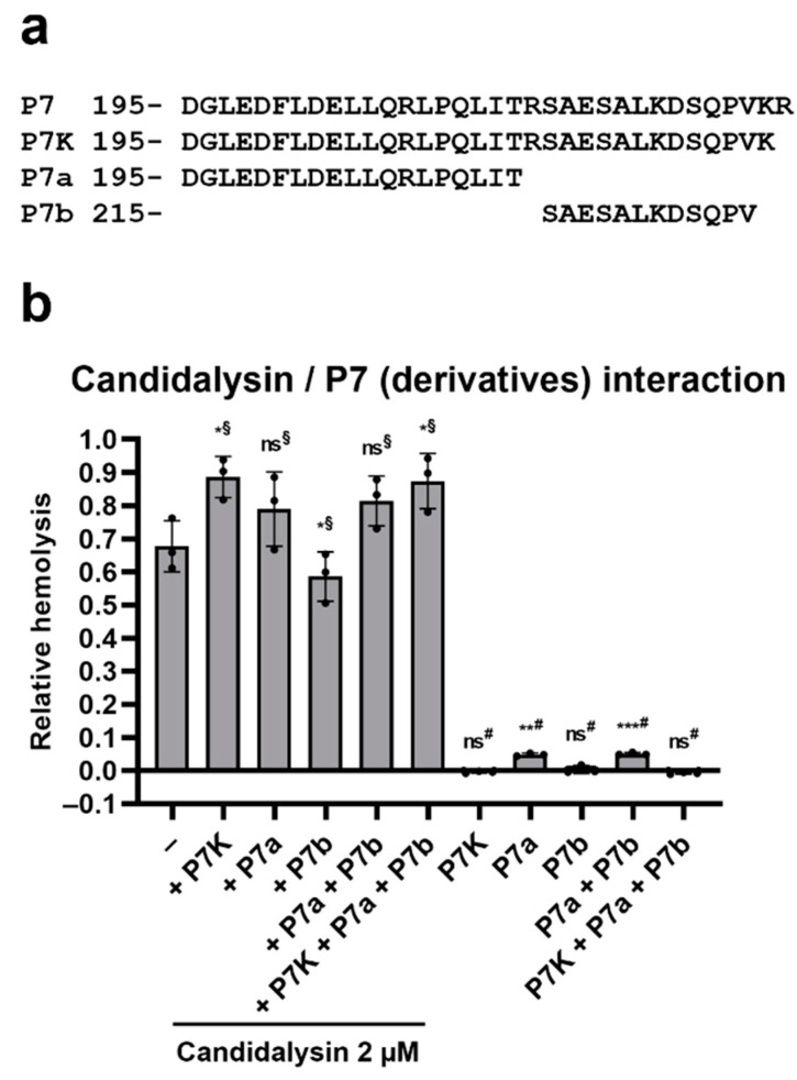
Figure 4.
P7 derivatives modulate the hemolytic potential of candidalysin. (a) Amino acid sequences of P7-derived peptides. Numbers represent the position of the first amino acid respective to the N-terminus of Ece1. (b) Purified RBCs were incubated with candidalysin and P7K, P7a and P7b (each peptide at 2 µM, Peptide Protein Research Ltd.) for 1 h at 37 °C. Hemolysis was quantified by measuring the absorbance of sample’s supernatant at 414 nm, and plotted relative to the full lysis control sample (RBCs incubated with pure water), following subtraction of the vehicle control. Each data point on the graph represents a different donor (average of 2 technical replicates). Error bars show the standard deviation. For statistical analysis, an arbitrary value of 0.01 was assigned to any value that was below this threshold. Student’s paired t-tests were then performed on log-transformed data. §, t-test vs. candidalysin only; #, t-test vs. vehicle only. *, p < 0.05; **, p < 0.01; ***, p < 0.005; ns, not significant.

