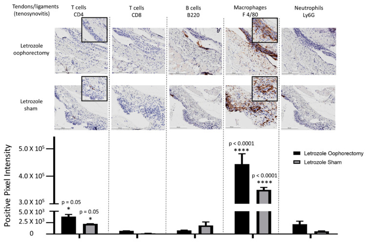
Figure 5.
IHC for immune cell subtypes in tenosynovitis infiltrates shows a macrophage-mediated inflammatory response induced by aromatase inhibitor treatment. Following oophorectomy or control (sham) surgery and induction of AIIA with daily letrozole injections, mouse legs were collected and processed for histopathological assessment as described in the Materials and Methods Section. To evaluate immune cell subtypes in the tendons, IHC staining of CD4+ T cells, CD8+ T cells, B220+ B cells, F 4/80+ macrophages, and Ly6G+ neutrophils was performed. Data were analyzed via two-way ANOVA for group-wise comparisons relative to each IHC stain. Values are the mean ± SEM, with indicated p values determined following Tukey’s corrections for multiple comparisons to measure the statistical change between IHC cell stain/subtype. * p ≤ 0.05; **** p ≤ 0.0001. All values of p ≤ 0.05 were considered statistically significant compared to all other stains individually.

