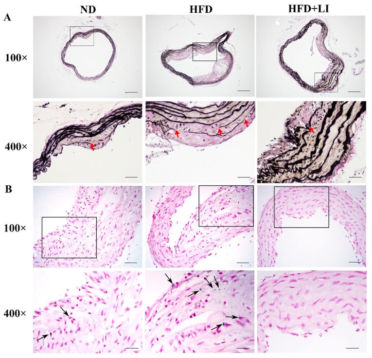
Figure 3.
Characterization of aortic elastic fibers and iron deposition in ApoE KO mice according to each diet group. (A) Voerhoff von Gieson staining of mice aortic tissues. (B) Prussian blue staining of murine aortic tissue sections assessed iron deposition in atherosclerotic plaques (n = 4). Arrowheads indicate positive staining areas. Images are shown at 100× and 400× magnification. Scale bar for 100× = 200 μm; scale bar for 400× =50 μm. ND: normal diet, HFD: high-fat diet, HFD + LI: high-fat diet without iron supplementation.

