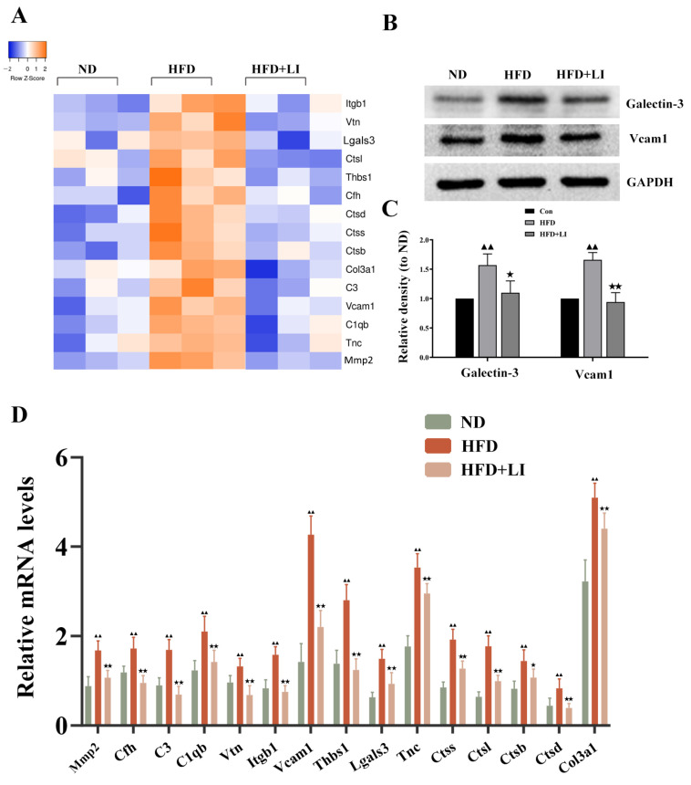
Figure 7.
Validation of the differentially expressed proteins identified with the iTRAQ analysis and involved in aortic inflammation, vascular remodeling, and focal adhesion. (A) Heatmap of inflammation, vascular remodeling, and focal adhesion-related protein expression in aortic tissues in each diet group. (B) qRT-PCR analysis of the protein expression in aortic tissues in each diet group (n = 4). Data is presented as mean ± SD. Samples were pooled from three independent experiments. (C) Western blot detection of Galectin-3 (gene name: Lgals3) and VCAM1 protein expression in aortic tissue. (D) Quantification of protein expression presented as density relative to GAPDH.▲▲ p < 0.01 vs. ND; ★ p < 0.05 vs. HFD; ★★ p < 0.01 vs. HFD. ND: normal diet-fed ApoE KO mice, HFD: high fat diet-fed ApoE KO mice, HFD + LI: high fat diet without iron supplementation-fed ApoE KO mice.

