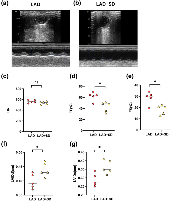
FIGURE 2.

Sleep deprivation deteriorated the left ventricular function of LAD mice. (a,b) Echocardiography results from mice in the LAD and LAD+SD groups. (c) The comparison of the HRs of mice in the LAD and LAD+SD groups is shown. (d) The comparison of the EF of mice in the LAD and LAD+SD groups is shown. The significance of differences between groups was determined using Welch's t‐test (n = 5, *p < 0.05). (e) The comparison of FS in mice from the LAD and LAD+SD groups is shown. The significance of differences between groups was determined using Welch's t‐test (n = 5, *p < 0.05). (f) The comparison of the LVIDd of the LAD and LAD+SD groups is shown. The significance of differences between groups was determined using Welch's t‐test (n = 5, *p < 0.05). (g) The comparison of the LVIDs in mice from the LAD and LAD+SD groups is shown. The significance of differences between groups was determined using Welch's t‐test (n = 5, *p < 0.05)
