
FIGURE 2.
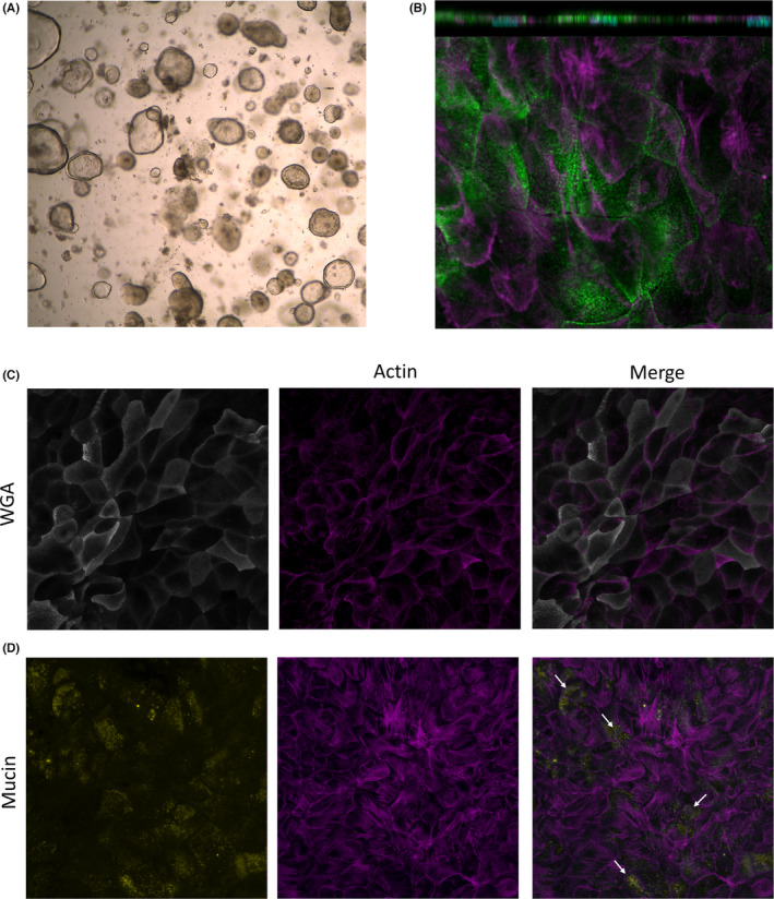
Characterization of differentiated colonoid monolayers. Colonoids obtained from colonic biopsies from a healthy subject were seeded as 2D‐monolayers and differentiated. (A) Image from light microscopy of BME‐suspended 3D colonoids; 4× magnification. (B) Polarized apical surface of cells defined by phospho‐ezrin (green) in differentiated monolayers. Orthogonal view (XZ projection), top panel. Top view (XY projection), bottom panel. (C) Glycocalyx‐rich cell membrane (wheat germ agglutinin, WGA in gray) and (D) mucin‐producing goblet cells (mucin in yellow) in differentiated colonoid monolayers. Mature microvilli and cell membrane borders on the apical surface were identified by actin expression (magenta). Nuclei are shown in cyan. Images were acquired with (A) Leica DM IL LED Inverted Microscope with ICC50 HD Camera and (B–D) LSM700 inverted confocal microscope; 20× magnification. BME, basement membrane extract