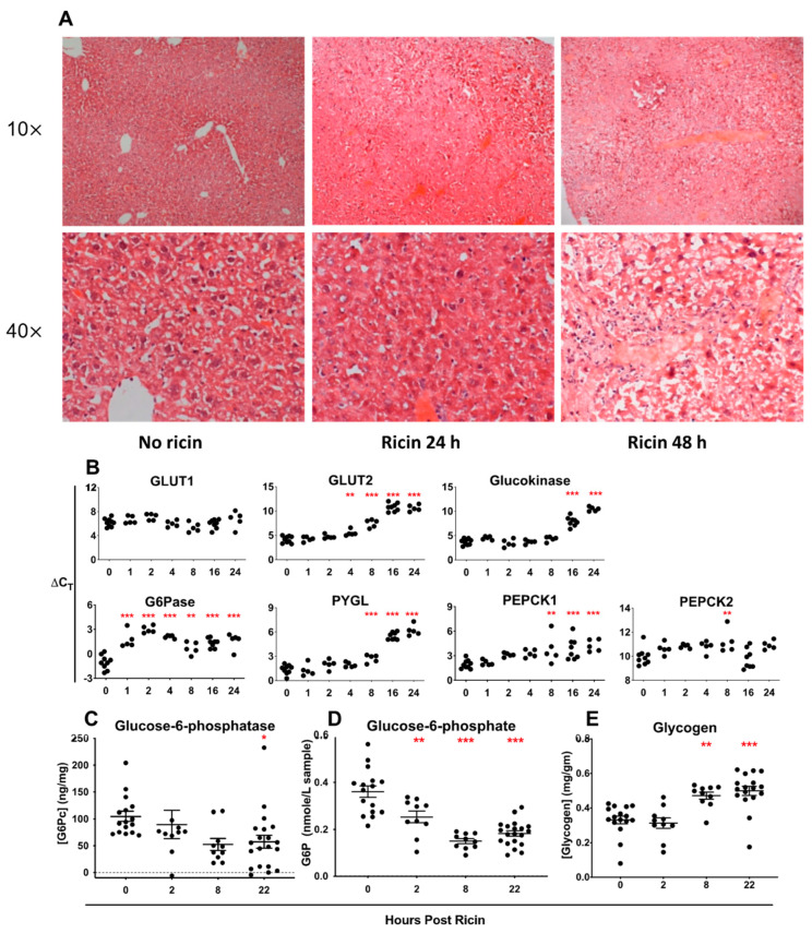
Figure 10.
Histochemical, gene expression, and biochemical profiles in the liver post ricin injection. The mice were injected with 30 µg/kg of ricin and sacrificed at indicated timepoints. The livers were removed and either immediately frozen or fixed in formalin. (A) Fixed tissues were stained with H&E and are shown at 10 and 40 magnification. (B) QRT PCR was performed on RNA extracted from the liver. ΔCt was calculated as described previously. GLUT: glucose transporter, G6Pase: glucose-6-phosphatase, PYGL: glycogen phosphorylase for liver, PEPCK: phosphoenolpyruvate carboxykinase. (C) ELISA was used to quantify the amount of the catalytic component of G6Pase present in BALB/c mice. (D) Glucose-6-phosphate was quantified in BALB/c mice using an enzyme assay. (E) Coupled en zyme assay was used to quantify glycogen in BALB/c mice. In panels (B–E), the Kruskal–Wallis test was performed and the red asterisks above the groups show the results of Dunn’s multiple comparison test vs. the control group: 0.05 (*), 0.002 (**), and <0.001 (***).

