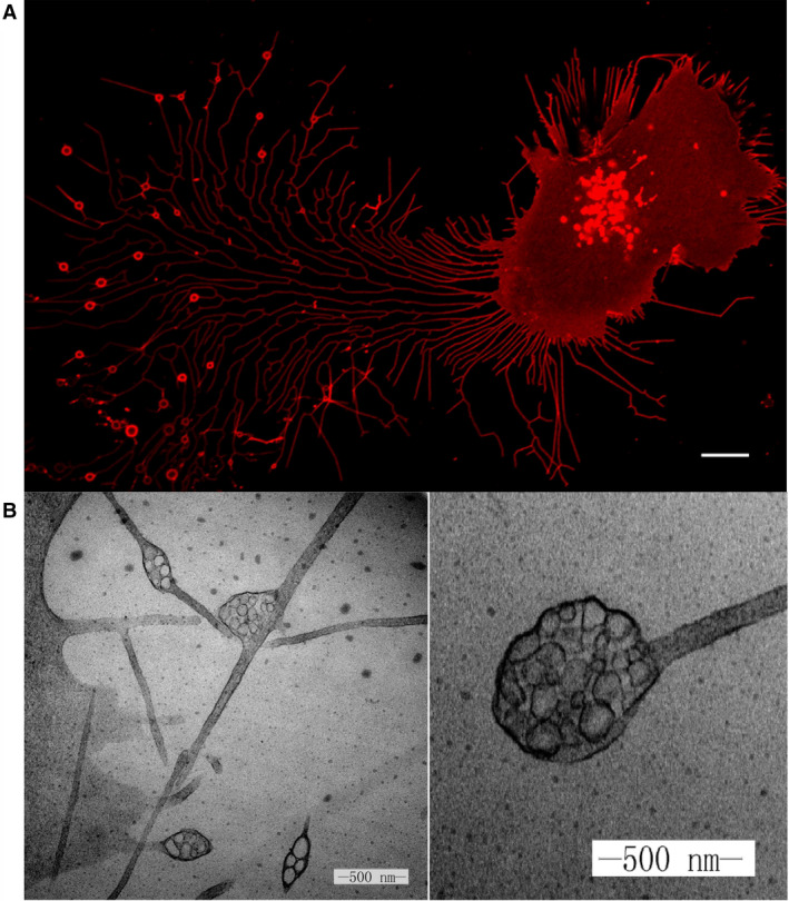
Fig. 1.

(A) Migrasomes from L929 cells. L929 cells were transfected with Tspan4‐mCherry and visualized by confocal microscopy. Scale bar, 10 mm. (B) Transmission electron microscope image of the pomegranate‐like structures, which we later named migrasomes. Scale bar, 500 nm.
