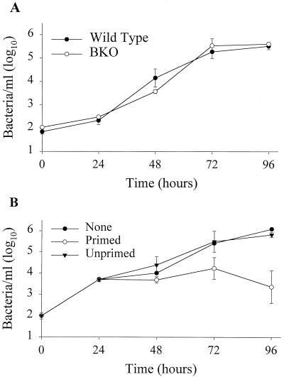
FIG. 1.
Growth and control of LVS in BMMφ. (A) Growth of LVS in wild-type and BKO BMMφ. BMMφ from wild-type and BKO mice were infected with LVS at an MOI of 1:10 (bacterium-to-macrophage ratio). At the specified time points after infection, BMMφ were washed, lysed, and plated. Data points show the mean numbers ± the standard error of the mean (SEM) of viable bacteria (triplicate samples) recovered from wild-type (filled circles) and BKO (open circles) BMMφ at the indicated time points. (B) Control of LVS growth by LVS-primed splenocytes. Uninfected mice (Unprimed) or mice primed intradermally with LVS 4 weeks prior to the onset of the experiment (Primed) served as the source of LVS-primed splenocytes. Immediately following LVS infection of BMMφ, 5 × 10⁶ splenocytes of each population were added to designated wells. At the indicated time points, cultures were assessed for intracellular bacteria. Data points show the mean numbers ± the SEM of viable bacteria (triplicate samples) recovered from macrophages alone (None, filled circles), cultures containing primed splenocytes (open circles), or cultures containing unprimed splenocytes (filled triangles) at the indicated time points; the SEM was too small to be visualized for some of the data points on this graph. The data in panel A are representative of three experiments similar in design, and those in panel B are representative of five experiments similar in design.

