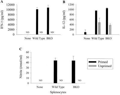
FIG. 6.
Secretion of cytokines and NO into culture supernatants following LVS infection. Splenocytes from either unprimed wild-type or BKO mice (gray bars) or mice primed with 8 × 10⁴ LVS 4 weeks prior to the onset of the experiment (black bars) were added to cultures of LVS-infected BMMφ. Culture supernatants (triplicate samples) were tested for IFN-γ (A), IL-12 (B), or NO (C) 72 h after addition of splenocytes. “None” represents LVS-infected BMMφ alone. ND, not detected. Each bar represents the mean ± the SEM cytokine or NO concentration of a group. These results are representative of three experiments similar in design.

