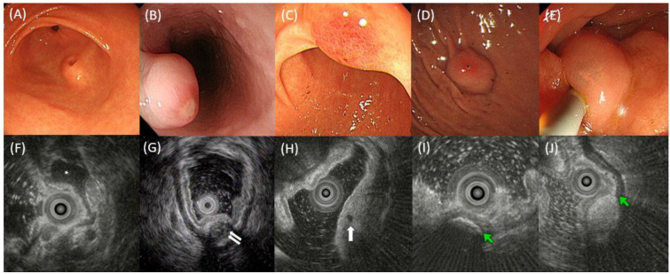
Figure 2.
Examples of the endoscopic and EUS findings of SELs. (A) Umbilication or central dimpling, (B) erosion or ulcer, (C) erythema, (D) hemorrhagic spot, and (E) translucidity. (F) EUS image showing a homogeneous hypoechoic SEL (asterisk) with a distinct border; (G) EUS image showing an SEL involving muscle propria layer (white arrows) with heterogeneity, mixed echogenicity, indistinct border, and deep attenuation; (H) EUS image showing anechoic foci (white arrow); (I) EUS image showing hyperechoic foci (green arrow) with acoustic shadowing that suggests calcification; (J) EUS image showing an SEL involving submucosal layer (green arrow indicates hyperechoic submucosal layer) with hyperechoic echogenicity, homogeneity, and distinct border. EUS, endoscopic ultrasonography; SEL, subepithelial lesion.

