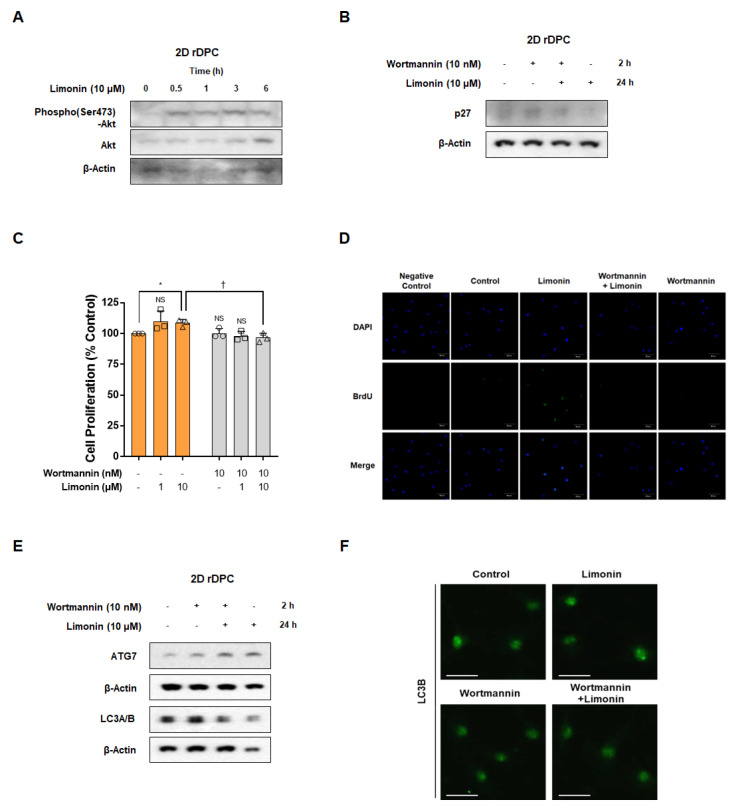
Figure 4.
Limonin induces cell proliferation through activation of the autophagy pathway via PI3K/AKT signaling in two-dimensional (2D) cultured rDPC. (A) Representative images of Western blots for phospho(Ser473)-AKT after treatment with limonin for 0–6 h. (B) Representative images of Western blots for p27 after treatment with limonin for 24 h in the absence or presence of wortmannin. (C) Changes in the proliferation of 2D dermal papilla cells after treatment with limonin for 3 days with or without wortmannin. (D) Confocal microscopy images for changes in the number of BrdU-positive cells after treatment with limonin for 24 h with or without wortmannin. (E) Representative images of Western blots for ATG7 and LC3A/B after treatment with limonin for 24 h with or without wortmannin. (F) Confocal microscopy images of changes in the number of LC3B puncta after treatment with limonin for 24 h with or without wortmannin. * p < 0.05 vs. vehicle-treated control. † p < 0.05 vs. limonin alone. Scale bars, 50 μm. NS, not significant.

