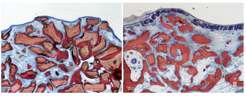
Figure 5.
Photomicrographs of ground sections representing the healing after 8 weeks in the submucosa region. Xenograft particles and soft tissues were present in both groups, but in higher percentages in the DBBM group. (A) DBBM group. (B) Collagenated group. Stevenel’s blue and alizarin red stain.

