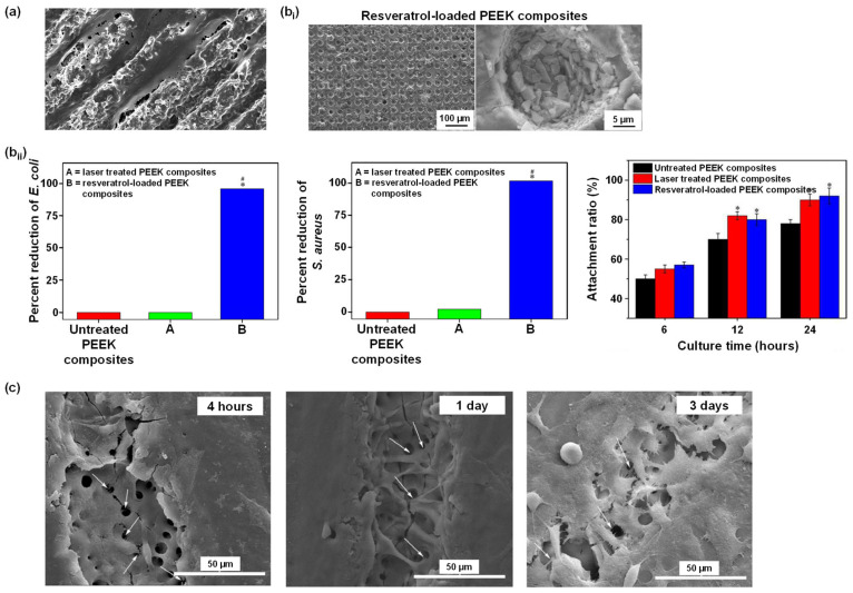
Figure 11.
Laser treatment of PEEK surfaces. (a) Gheisarifar et al. observed cell alignment and elongation following the geometry of the surface topography (grooves) [40]. (b) The work from Cai et al.: (bi) SEM images showed porous PEEK composites loaded with antibacterial agent resveratrol; (bii) antibacterial activity (* p < 0.05 against untreated PEEK, # p < 0.05 against laser treated PEEK) and cell attachment ratio (* p < 0.05 against untreated PEEK), respectively [205]. (c) SEM images showed the morphology of MC3T3-E1 pre-osteoblasts cultured for 4 h, 1 d, and 3 d. White arrows: cell pseudopodia protruding into the laser-generated pores [206]. All figures were re-printed with permission from the literature [40,205,206].

