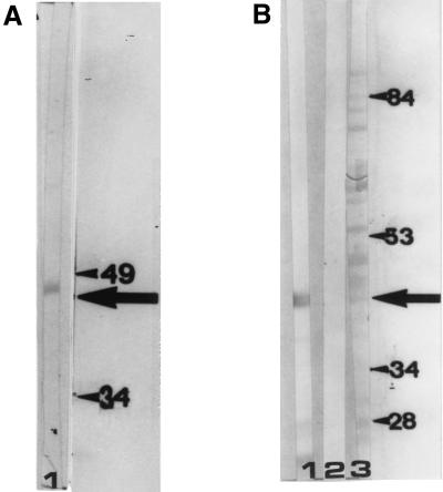
FIG. 2.
(A) Reactivity of human anti-p42 antibodies with rSG3PDH. A strip of induced bacterial extract was incubated with human anti-p42 antibodies in Western blotting (lane 1). The arrow indicates the migration position of rSG3PDH. On the right are molecular masses (in kilodaltons) of protein standards. (B) Reactivity of mouse anti-rSG3PDH antibodies with SAWA p42. Strips of SAWA blot were incubated with immune (lane 1) or control (lane 2) mouse serum in Western blotting or stained with Ponceau red (lane 3). The arrow points to SAWA p42. On the right are molecular masses (in kilodaltons) of protein standards.

