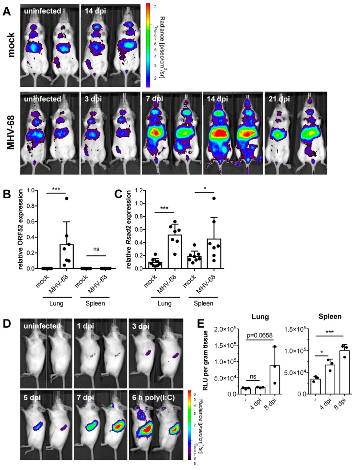
Figure 1.
Acute MHV-68 infection induces IFN-β and ISG expression in vivo. (A) IFN-β luciferase reporter mice were intranasally infected with 5 × 10⁴ PFU MHV-68. Mock-infected control mice received PBS only. Whole-body in vivo imaging was performed at the indicated days post-infection. Two representative mice of each group are shown (n = 3–5). (B,C) Wild-type mice were intranasally infected with 5 × 10⁴ PFU MHV-68 and sacrificed seven days after infection. Relative expression of MHV-68 ORF52 and of mouse Rsad2 in lung and spleen tissue was determined by qPCR (n = 7–8, mean ± SD). p-values were calculated by one-way ANOVA followed by Sidack’s Multiple Comparison Test. ns, p > 0.05; *, p ≤ 0.05; ***, p ≤ 0.001. (D) Whole-body in vivo imaging of luciferase activity of adoptively transferred Mx2Luc reporter splenocytes (1 × 10⁷ cells/mouse) upon intranasal infection of recipient mice with 5 × 10⁴ PFU MHV-68. Control mice were intravenously injected with 20 µg poly(I:C) and imaged after 6 h. Two mice from a representative experiment are shown. (E) In vitro analysis of luciferase activity in spleen and lung tissue four and eight days after intranasal infection of Mx2Luc reporter mice with 5 × 10⁴ PFU MHV-68 (n = 3, mean ± SD). p values were calculated by one-way ANOVA followed by Dunnett’s Multiple Comparison Test. ns, p > 0.05; *, p ≤ 0.05; ***, p ≤ 0.001.

