Figure 3.
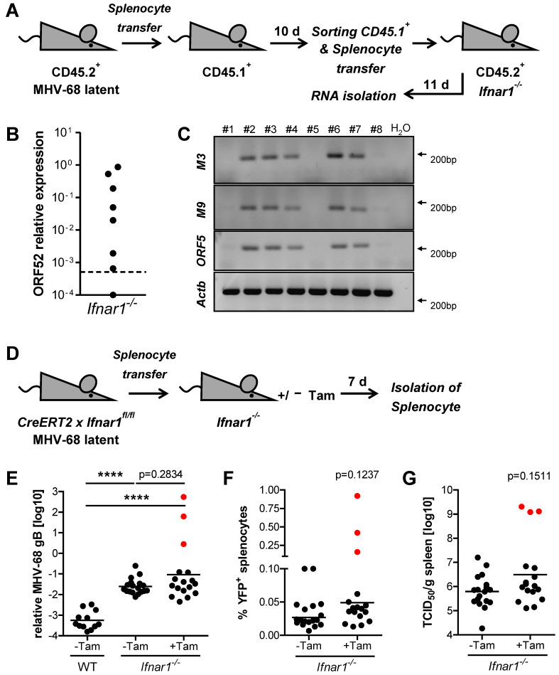
MHV-68 is transmitted during latency despite an intact type I IFN system. (A) Schematic of adoptive transfer model. Splenocytes were isolated and pooled from latently infected CD45.2+ mice (≥21 days after infection) and adoptively transferred (1 × 10⁷ cells/mouse) into CD45.1+ recipient mice (n = 8). Recipient mice were sacrificed after 10 days, and CD45.1+ splenocytes were purified by FACS and adoptively transferred (7.5 × 10⁶ cells/mouse) into Ifnar1−/− mice (n = 8). Total RNA was isolated from splenocytes 11 days after the transfer into Ifnar1−/− recipient mice. (B) Quantification of MHV-68 ORF52 expression by qPCR in splenocytes of Ifnar1−/− mice 11 days after adoptive transfer. The dotted line represents the level of relative ORF52 expression in pooled CD45.1+ donor splenocytes before adoptive transfer into Ifnar1−/− mice. (C) Conventional RT-PCR on MHV-68 transcripts M3, M9, and ORF52 in spleen tissue of Ifnar1−/− mice 11 days after the second adoptive transfer. (D) Schematic of adoptive transfer model. Splenocytes were isolated from latently infected (MHV-68 H2bYFP), tamoxifen-inducible Ifnar1−/− (R26CreERT2 × Ifnar1fl/fl) mice and adoptively transferred (1 × 10⁷ cells/mouse) into Ifnar1−/− recipient mice. Recipient mice were subsequently treated orally with (+Tam, n = 17) or without (-Tam, n = 18) a single dose of 2 mg tamoxifen. (E–G) Relative quantification of MHV-68 genomic DNA (gB) by qPCR, frequency of YFP+ splenocytes, and quantification of recoverable infectious by TCID50 assay in spleen tissue of recipient mice seven days after adoptive transfer (geometric mean is indicated). WT recipient mice received latently infected splenocytes in the absence of tamoxifen (n = 12). Red dots indicate measurements for the same three mice. p-values were calculated by Mann-Whitney’s U test. ****, p ≤ 0.0001.

