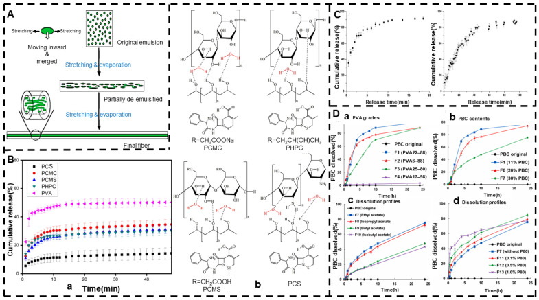
Figure 5.
Emulsion electrospinning. (A) Schematic diagram of the mechanism of core–shell fiber formation during emulsion electrospinning (reprinted with permission from [158]. Copyright©2006, WILEY-VCH Verlag GmbH & Co., KGaA, Weinheim). (B) (a) In vitro drug release of PVA/biopolymer mixtures and (b) intermolecular interactions between PVA and different biopolymers (reprinted with permission from [164]. Rights managed by Taylor & Francis). (C) In vitro drug release curve of PCL fibers containing KET and PCL/gelatin crosslinked fibers (reprinted with permission from [166]. Copyright©2017, Elsevier B.V. All rights reserved.). (D) (a–d) Release curves of PVA grade, PBC content, use of different solvents, effect of P80 concentration on PBC dissolution of PPA nanofibers (reprinted with permission from [170]. Copyright©2021, Elsevier B.V. All rights reserved).

