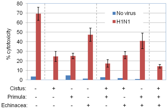
Figure 4.
Prophylactic effects of P. veris on influenza virus cytolysis. MDCK cells were incubated for 4 h with 50 μg/mL Primula extracts isolated by supercritical CO2 + 13% EtOH, and then infected with H1N1 at MOI = 0.2 according to the protocol of Figure 1B. Echinacea (EtOH/H2O-Xad7-derived) and Cistus (hydroethanolic) extracts were used in parallel for comparison, in the presence or absence of Primula extract. The graph depicts virus-induced MDCK cell lysis, assessed by LDH release assays. Data are the average of duplicate determinations from 3 independent experiments.

