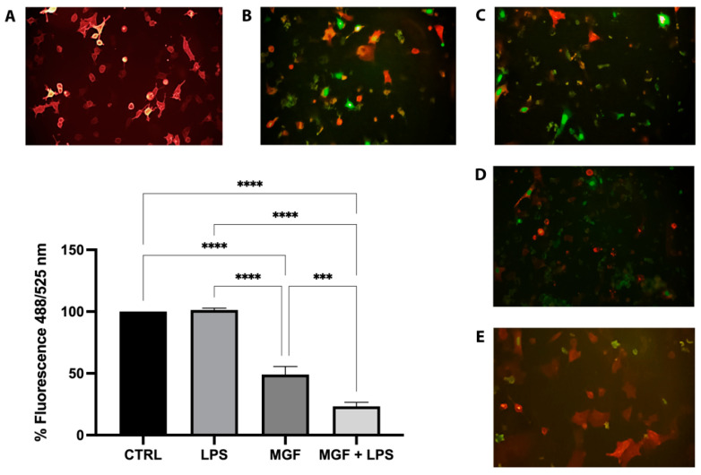
Figure 7.
Representative images of pseudo SARS-CoV-2 entry: entry of pseudovirus bearing the green reporter in red fluorescence ACE2-expressing host cells A549. Green fluorescence nucleus was only expressed in cells infected by the pseudovirus; therefore, the amount of green present was proportional to the number of infected cells. (A) K cells, A549 transduced with ACE2 BacMam; (B) K virus, cells infected by pseudo SARS-CoV-2; (C) LPS treated cells; (D) MGF treated cells; (E) co-treated MGF + LPS cells. Images obtained at 20X magnification. *** p < 0.001; **** p < 0.0001.

