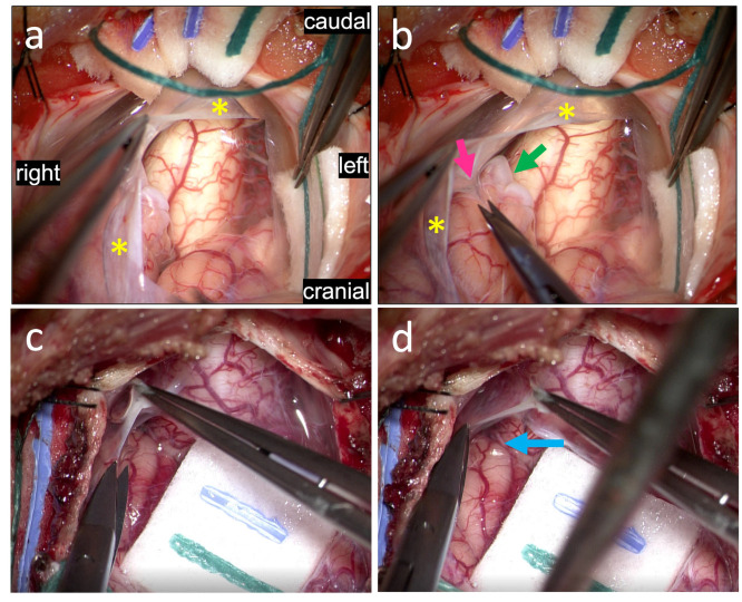
Figure 5.
View of the posterior craniocervical junction following durotomy. After flapping up the dura, the underlying arachnoid (yellow asterisk) can be visualized and is subsequently resected (a). The arachnoid is thickened in these areas and the underlying tonsils appear gliotic (green arrow), with numerous associated arachnoid adhesions (magenta arrow) that appear to tether the cerebellar tonsils to the arachnoid and overlying dura (b). These adhesions are carefully dissected out and cut (not shown). For comparison, a non-CM1 case is shown, with non-gliotic tonsils free from arachnoid tethering (c,d).

