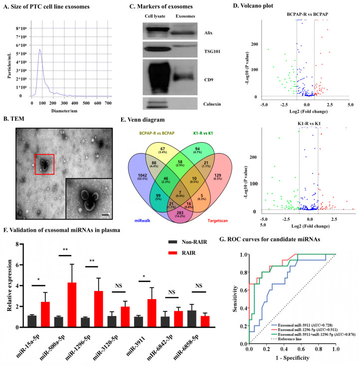
Figure 2.
Identification and verification of candidate exosomal miRNAs associated with RAIR PTC. (A). NTA results suggested that exosomes were about 50–150 nm in diameter. (B). TEM images showed that exosomes were oval or bowl-shaped capsules without the nucleus. The red flame is the representative image. scale bare, 100 nm (C). exosomes markers Alix, CD9 and TSG101 were all detected and Calnexin, a negative marker of exosomes was absent in our isolated exosomes. (D). Differential expressed exosomal miRNA in treated PTC and parental cell lines. (E). A Venn diagram showed differentially expressed exosomal miRNAs (RAIR cell lines vs. parental cell lines) and miRNAs related to the NIS (predicted by miRwalk and Targetscan). (F). Validation of the differentially expressed selected plasma exosomal miRNAs in PTC patients with non-131I-avid metastases and 131I-avid metastases. (G). Receiver operating characteristic (ROC) curve of exosomal miR-1296-5p, miR-3911 and their combinations as a predictive marker for radio-iodine refractory PTC. NS, no significance, * p < 0.05, ** p < 0.01.

