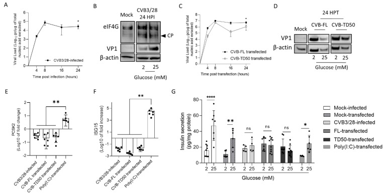
Figure 5.
Infection by CVB3/28 and transfection by CVB-TD50 RNA forms decrease PCSK2 mRNA expression and abolish the insulin response to glucose stimulation in beta cells of rodents. (A) Infectious CVB3/28 virus loads in the supernatant of INS-1 cells at indicated time points post-infection. (B) Western blot analyses showed the cleavage of eIFG4, a eukaryote translation factor, viral protein 1 (VP1) and β-actin expression in mock-infected and CVB3/28-infected pancreases at 24 h post-infection (24 HPI) in INS-1 cells. (C) Kinetics of viral RNA replication activity after CVB-FL and CVB-TD50 RNA forms (1 μg) transfection of INS-1 cells measured by RT-qPCR. (D) Western blot analysis of CVB3/28 viral protein 1 (VP1) and β-actin in mock-infected and CVB-FL/TD50-transfected INS-1 cells at 24 h post-transfection (24 HPT). (E) PC2 (PCSK2) mRNA level fold-changes (RT-qPCR) in INS-1 cells infected with CVB3/28, or transfected with CVB-FL/TD50 or Poly (I:C). (F) ISG15 mRNA level fold-changes (RT-qPCR) in INS-1 cells infected with CVB3/28, or transfected with CVB-FL/TD50 or Poly (I:C). (G) Glucose stimulated the insulin secretion assay of INS-1 cells following infection of CVB3/28, transfection of CVB RNA forms or Poly (I:C), or mock-infected/transfected cells (triplicate). Data are expressed as pg of insulin in supernatant per mg of total protein content. ANOVA test (panel (G)) or Mann–Whitney U test (panels (A,C,E,F)); *: p < 0.05; **: p < 0.01; ****: p < 0.0001. Not specified or ns: non-significant. Data represent mean +/− SD of three independent experiments. CVB-FL/TD: full-length or 5′ terminally deleted coxsackievirus. HPI: hours post-infection. HPT: hours post-transfection. CP: cleavage product. VP1: viral capsid protein 1. eIF4G: Eukaryotic translation initiation factor 4G.

