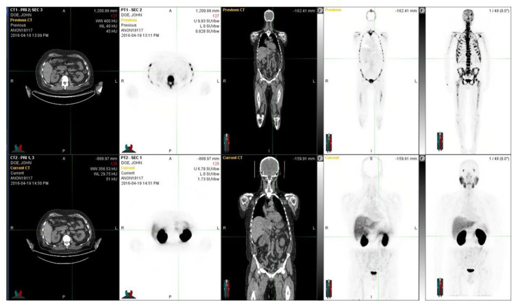
Figure 2.
The figure demonstrates a 60-year-old prostate cancer patient diagnosed five years earlier with prostate cancer and T3bN0M0 with an initial PSA 13 and a GS 7. Despite the radical prostatectomy and hormonal therapy, he developed skeletal metastases. He was treated with chemotherapy and multiple targeted bone therapies, abiraterone, and Ra-223 radionuclide therapy. In the upper row, NaF-PET demonstrates a widespread skeletal disease in the whole skeleton, whereas in the lower row, with Ga-PSMA, fewer skeletal metastases are seen. Regions differed significantly from each other, and the PET tracers showed different overall distributions. Fluoride typically targets the cortical bone and bone formation, whereas PSMA targets active cancer cells, preferably in the bone marrow. At the time of imagings, the S-PSA was 0.35 (patient 7).

