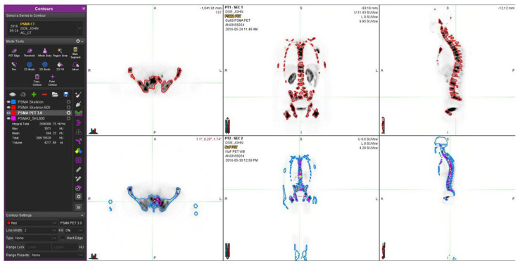
Figure 5.
A 66-year-old patient with a T4N1M1 (GS 8) prostate cancer diagnosed three years earlier with an initial PSA of 15.0 is shown. He was originally treated with androgen deprivation and chemotherapy. After relapses, multiple new targeted bone therapies were introduced: abiraterone, Ra-223, and denosumab. At the time of the investigation, widespread skeletal disease (not very active) was observed both with NaF PET (lower row) and Ga-PSMA PET (upper row). There were essential differences in their distributions, as seen especially in the transaxial images. Fluoride typically targets the cortical bone and bone formation, whereas PSMA targets the active cancer cells, preferably in the bone marrow. Here, in the NaF images, the skeleton has better recovered from the targeted treatment, as the bone marrow still shows some activity. At the time of imaging, the S-PSA was 3.6 (patient 3).

