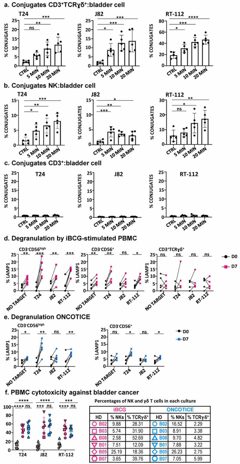
Figure 6.

Effector function of PBMC after 7-day exposure to BCG.
PBMC from healthy donors were incubated with iBCG or Oncotice as indicated. After 7 days in culture, cells were recovered for their use as effector in functional assays to enquire the recognition and killing of target cells (the bladder cancer cell lines T24, J82, RT-112; K562 cells were used as NK positive control target). a, b, c. Conjugate formation between effector and target cells. BCG-treated cells were incubated for 5–20 min with CellTraceTM Violet-dyed target cells at 37°C (except negative control, kept at 4°C). Conjugate formation was measured for double T-cells stained by lymphocyte markers (either CD3+TCRγδ+, CD56+ for NK cells or CD3+TCRγδ− for CD3+, presumably TCRαβ) and violet dye. Statistical analysis was performed using one-way ANOVA comparing each condition with the untreated culture (*, p < .05; **, p < .01; ns, non-significant). d, e. Degranulation. The functional capacity of activated NK cells, as shown by CD56 upregulation (CD56high) and the rest of the CD3−CD56+ NK cells (CD56+) was analyzed separately, as indicated, as well as degranulation activity of γδ T-cells (in Oncotice-treated cells this population was scarce, so its analysis is not shown). 25,000 effector cells (normalizing for NK cells) were incubated with 50000 target cells (1:2 E:T ratio) and surface LAMP-1 (CD107a) was measured. Statistical analysis was done by one-way ANOVA. f. Specific cytotoxicity. Effector cells were included in cytotoxicity assays against target cells labeled with calcein-AM. Each symbol represents a different healthy donor (HD). Percentages of NK and γδ T-cells in each culture are included in a table (right). Pink: iBCG; Blue: Oncotice. Statistical analysis was done by one-way ANOVA.
