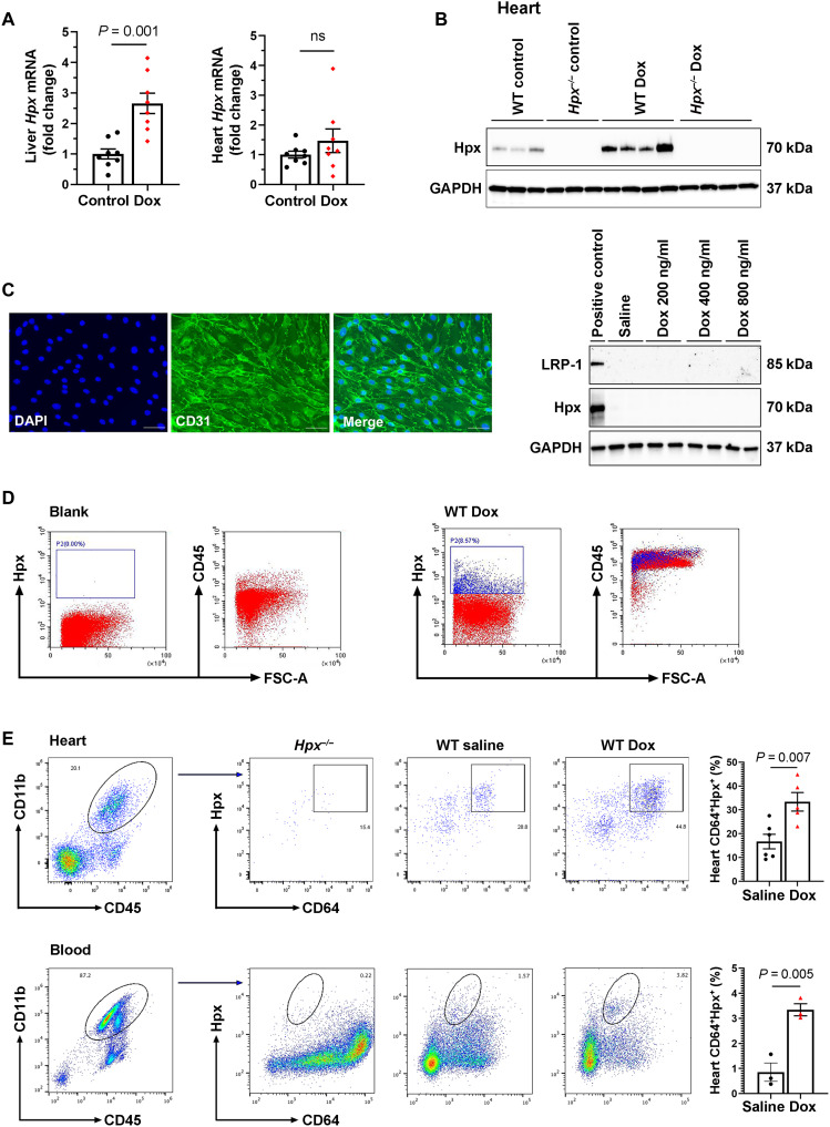
Fig. 6. Hpx may be transported to the heart by circulating macrophages.
(A) Hpx mRNA levels in the liver and heart in mice treated with saline (control; n = 8) and Dox (n = 8) mice within 24 hours after completion of the 2-week regimen. (B) Cardiac Hpx protein level within 24 hours after completion of the 2-week regimen (n = 8). (C) Glyceraldehyde-3-phosphate dehydrogenase (GAPDH), Hpx, and LRP-1 detected by Western blot in adult MHECs treated with saline (control) or Dox at a concentration of 200, 400, and 800 ng/ml for 48 hours. The left-most lane is a liver sample that was used as a positive control. DAPI, 4′,6-diamidino-2-phenylindole. (D) Circulating Hpx-expressing cells were CD45+ immune cells, as determined by fluorescence-activated cell sorting (FACS). FSC-A, forward scatter area. (E) Hpx-expressing cells were CD11b+CD64+ macrophages in the heart and circulation as determined by FACS. Data were expressed as means ± SEM. Welch’s t test was used to compare the difference between control and Dox-treated groups.

